Chest imaging

chest imaging

Emphysema

Emphysema is characterized by permanently enlarged airspaces distal to the terminal bronchiole, with destruction of alveolar walls. Emphysema is usually classified according to the part of the acinus predominantly affected. The Computed Tomography appearance of emphysema consists of focal areas or regions of low attenuation, usually without visible walls.

chest imaging

Airway disease - Bronchial Wall Thickening

Bronchial wall thickening is an imaging descriptor used to describe abnormal thickening of bronchial walls and can arise from a vast number of pathological entities. It is one of the causes of peribronchial cuffing. The presence of bronchial wall thickening usually implies inflammation of the airways.

chest imaging

Airway disease - Bronchiectasis

Irreversible localized or diffuse bronchial dilatation, usually resulting from chronic infection, proximal airway obstruction, or congenital bronchial abnormality.

chest imaging

Airway disease - Centrilobular nodules

Rounded or irregular opacity, well or poorly defined, measuring up to 3 cm in diameter. Centrilobular nodules appear separated by several millimeters from the pleural surfaces, fissures, and interlobular septa. They usually are ill-defined, with soft-tissue or ground-glass attenuation, and size ranging from a few millimeters to one centimeter.

chest imaging

Airway disease - Air trapping

Air trapping represents the retention of air in the lung distal to an obstruction. Air trapping is best evaluated on end-expiration CT scans as parenchymal areas with less than normal increase in attenuation and lack of volume reduction. Comparison between inspiratory and expiratory CT scans is helpful when air trapping is subtle or diffuse. Differentiating air trapping from lung parenchymal mosaic perfusion is a major challenge when interpreting end-inspiratory CT images.

chest imaging

Lung fibrosis

Lung fibrosis can be qualitatively graded into three levels: 0-10%, 11-30%, and > 30%. Usual interstitial pneumonia (UIP) and nonspecific interstitial pneumonia (NSIP) are the main CT patterns of lung fibrosis.

chest imaging

Centrilobular ground-glass nodules

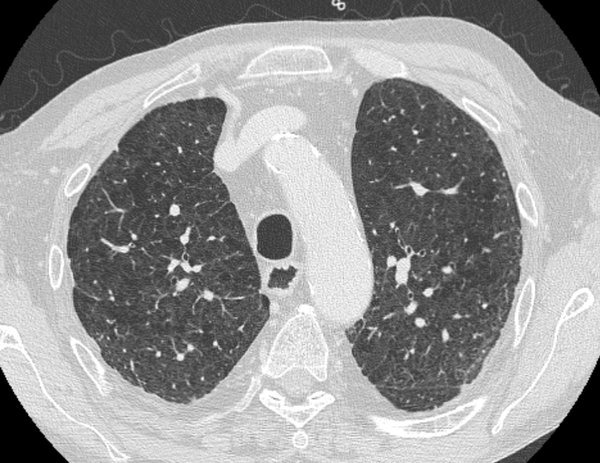
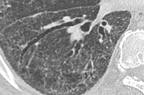
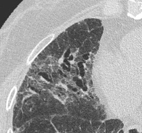
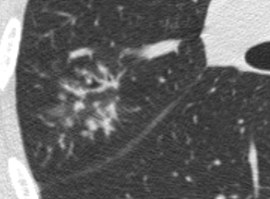
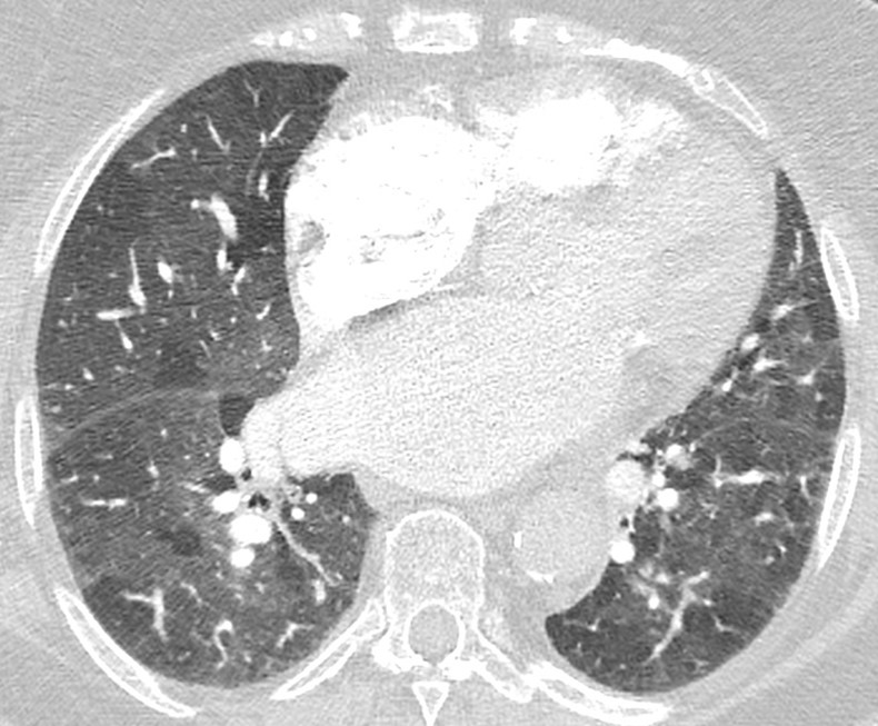
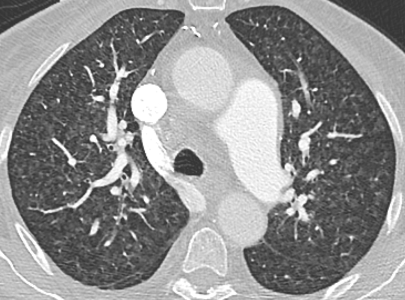
Nodules with centrilobular distribution locate a few millimeters away from the pleural surface and fissures, without direct contact with them. Diffusely distributed, poorly defined centrilobular ground-glass nodules are commonly observed in patients with idiopathic PAH. These nodules correspond to cholesterol granulomas resulting from the ingestion of red blood cells by pulmonary macrophages. The differential diagnosis includes hypersensitivity pneumonitis, respiratory bronchiolitis, follicular bronchiolitis, and long-standing left-to-right shunt. It is worth noting that centrilobular ground-glass nodules, along with tortuous corkscrew vessels, can also be observed in pulmonary capillary hemangiomatosis (PCH) and pulmonary veno-occlusive disease (PVOD).

chest imaging

Mosaic attenuation pattern

This pattern appears as patchwork of regions of differing attenuation that may represent (a) patchy interstitial disease, (b) obliterative small airways disease, or (c) occlusive vascular disease. CT features suggesting vascular origin include signs of primary PH, chronic pulmonary embolism, and PVOD.

chest imaging

Left atrium enlargement

The diameter measurements of the left atrium on CT axial images should be determined as the largest anterior-posterior diameter. It is a continuous, metric variable.

chest imaging

Coronary artery calcification

Coronary artery calcification can be identified as high-attenuation material in the path of a coronary artery. For non-gated thoracic CT in routine clinical practice, it is recommended to use a simple visual ordinal score performed on a whole-patient basis. CAC is scored as absent, mild, moderate, or severe on a whole-patient basis to summarize the cumulative findings in all the coronary arteries. This approach can be applied to both non-contrast and contrast-enhanced images.

chest imaging

Valvular calcification

Valvular calcification can be identified as high-attenuation material in the region of the aortic valve. Care should be taken to differentiate valve calcification from calcification in the aortic root, mitral annulus, or coronary arteries (aortic valve).

chest imaging

Congenital heart disease

The pathophysiology of congenital heart disease is variable and complex, affecting management decisions. In patients with large shunts, pulmonary vascular disease develops secondary to shear stress, endothelial damage, smooth muscle proliferation, remodeling of the vasculature, and ultimately obliteration of the microvasculature, similar to idiopathic PAH.

chest imaging

Signs of portal hypertension

Presence of enlarged portosystemic collateral vessels in the chest and upper abdomen. Coronary venous collaterals are typically associated with esophageal or paraesophageal varices. Esophageal varices are commonly fed by the anterior branch of the left gastric vein, while the posterior branch of this vein supplies paraesophageal varices. On CT, varices are distinct, round, tubular, or winding structures that exhibit smooth, homogeneous attenuation and enhance with contrast material to a similar degree as neighboring portal and mesenteric veins.